On a peaceful afternoon in early summer 1977, the laboratory of CERN radiobiologist Marilena Bianchi was visited by a physicist with a pretty unusual request. He asked for her help in his quest to create a first image of a mouse using a PET (positron-emission tomography) camera.
The physicist, David Townsend, had been helping Alan Jeavons, also a physicist at CERN. Jeavons had developed a new detector, based on a high-density avalanche chamber, to take PET images. Townsend had developed the software to reconstruct the data from the detector and to turn them into an image.
Once they were ready, Townsend asked Bianchi, who was developing medical applications of CERN technologies, to inject a mouse with a small amount of short-lived radioisotope, which was absorbed into the skeleton of the animal.
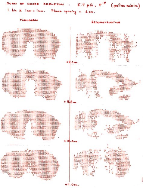
The isotope she injected emitted positrons, the antimatter twins of electrons. These positrons bumped into nearby electrons and in the collision a pair of photons was created. The photons shot out in exactly opposite directions. By placing two detectors around the mouse, Jeavons and Townsend picked up these pairs of photons, pinpointing where the positron annihilations occurred. “A few days later, David Townsend came back with this beautiful picture. The first mouse scan taken with a PET camera,” remembers Streit-Bianchi. “The findings were then presented at a conference in October 1977.”
PET was not invented at CERN, but the work carried out by Jeavons and Townsend made a major contribution to its development, thanks to the type of detector and computer programme developed for image-taking analysis. After the initial success, Jeavons and Townsend devoted their careers to improving medical imaging. Later, Townsend and co-workers in the US suggested to combine PET-CT (computed tomography) to see both metabolic and anatomic information. This was a major breakthrough for cancer diagnosis and treatment follow up.
“I am very proud. The inventiveness of these two physicists and their desire to develop a special PET camera resulted in the further development of a perfectly safe method to inquire what is happening in the body.” - Streit-Bianchi
Forty years on, PET technology is even more advanced thanks to the work carried out at CERN and other research laboratories around the world. The technologies and scientific advances behind high-energy physics – through developments in accelerators, detectors and computing – have helped to contribute to the field of medical imaging.
Twenty years ago, with Marilena Streit-Bianchi’s help, CERN established a dedicated policy and structure for knowledge and technology transfer. The CERN group of the Crystal Clear Collaboration is now developing new fast detector prototypes for use in both high-energy physics experiments and medical imaging, with particular emphasis on the PET technology. Read more about Streit-Bianchi’s 41-year career at CERN in the September 2014 CERN Courier.
Find out more about PET at CERN in the June 2005 CERN Courier.